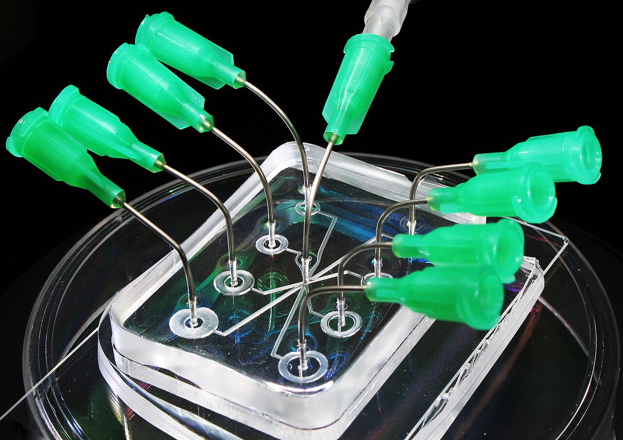
A microfluidic chip developed by scientists at Southern Federal University and their international colleagues is designed to isolate rare circulating tumor cells in the blood that can initiate metastasis in different organs. The study’s findings were published in Microchemical Journal.
Team in Russia isolates cells from the bloodstream
The device, described as being about the size of a coin, targets folate receptors, which are more abundant in cancer cells than in healthy cells. The chip’s channels are coated with a film based on chitosan, a natural polymer obtained from crustaceans. Folate molecules and aptamers—synthetic DNA fragments—serve as structures on this surface that capture tumor cells.
As blood moves through the device’s labyrinth-shaped channels, cancer cells attach to the channel walls while other cells pass through with the flow. This is intended to enable rare tumor cells to be isolated alive from several milliliters of blood in about 20 minutes.

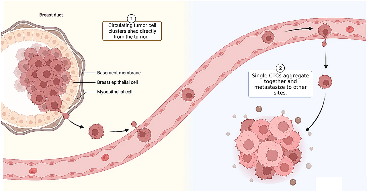
Metastasis occurs when individual cancer cells that have broken away from the primary tumor reach other tissues through the blood or lymphatic system. Their scarcity among billions of blood cells makes them difficult to detect.
Chip captures 80 to 91 percent of cells in breast tumors
At this stage of the study, the chip was adapted to identify cells from breast tumors. The experiments captured 80 to 91 percent of circulating tumor cells, while no false-positive results were observed in blood samples from healthy individuals.
More than 95 percent of the captured cells remained alive and undamaged. This could enable the cells to be examined later at the molecular level and the disease to be monitored through a routine blood test.
Chip could be adapted for different types of cancer
The new device can be produced with a 3D printer in about 45 minutes, and the molecular structures used can be changed according to the type of cancer. The researchers aim for this approach to make the technology and device more economically accessible.
The study also included flow cytometry, magnetic resonance imaging and computed tomography among existing diagnostic applications. It noted that some immune-based magnetic and microfluidic systems can result in cell loss or damage when cells are manually transferred to another section for analysis.
First stage before clinical trials
The results were described as the first stage before clinical trials. While the team at Southern Federal University is working on new microfluidic device geometries, researchers in Krasnoyarsk are focusing on increasing the selectivity of cell capture with aptamers.
Researchers from Krasnoyarsk State Medical University, the Krasnoyarsk Science Center of the Siberian Branch of the Russian Academy of Sciences, the Krasnoyarsk Regional Clinical Oncology Dispensary and Punjab University in India also took part in the project. The study was conducted under Southern Federal University’s “Accelerated production of micro- and small-tonnage materials” project and the “Priority-2030” strategic academic leadership program, with support from Russia’s Ministry of Health and Ministry of Science and Higher Education.