- BIST10010,760-3.63%
- USD49.86+0.15%
- EUR55.82+0.52%
- GOLD3,898.90-3.29%
- INTEREST36.00%-1.00
- BITCOIN77,712+0.35%
- November 1, 202614°
- BIST10010,760-3.63%
- USD49.86+0.15%
- EUR55.82+0.52%
- GOLD3,898.90-3.29%
- INTEREST36.00%-1.00
- BITCOIN77,712+0.35%
- November 1, 202614°
Moscow surgeons seal 13-year-old’s faulty lymphatic vessels with clips
At Moscow’s Morozov Hospital, doctors sealed the faulty lymphatic vessels of a 13-year-old with a congenital lymphatic vessel disorder that caused fluid to accumulate in the lungs. Following what was the first operation of its kind in pediatric healthcare in the city, the child was discharged and returned to normal life.


Doctors surgically treated a congenital lymphatic vessel disorder at Moscow’s Morozov Hospital that had caused fluid to accumulate in a 13-year-old’s lungs. Using specialized imaging techniques, they located the faulty lymphatic vessels and sealed them with clips.
The disorder was detected during fluorography
Fluid in the child’s lungs was discovered during fluorography performed as a precautionary screening. Hospital examinations subsequently identified a congenital lymphatic vessel disorder.
The disease had previously caused no symptoms. Because the lymphatic system had developed abnormally, lymph leaked into and accumulated in the chest cavity. The condition gradually weakened the body without being noticed and is known in medical literature as chylothorax.
Lymphatic pathways located with a fluorescence camera
Pavel Mordvin, head of the surgical department, said the lymphatic vessels were less than 1 millimeter thick and almost invisible to the naked eye. He said this made the operation highly demanding in terms of precision.
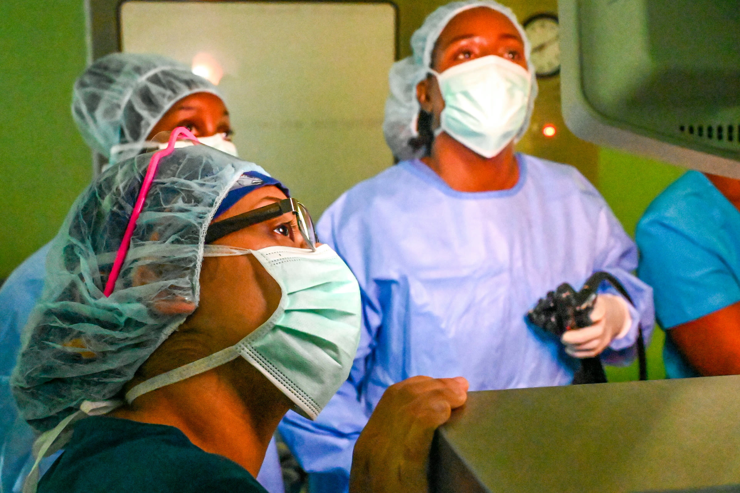
Surgeons injected a dye called indocyanine green through small incisions in the abdomen, making the lymphatic pathways visible. They then used a fluorescence camera through small incisions in the chest to locate the faulty vessels.
The identified lymphatic vessels were sealed with clips. The operation was the first use of this method in pediatric healthcare in Moscow.
The child left the hospital
Following the procedure, the child was discharged from the hospital and returned to normal life.
Stay Updated with RuskiGazeta
Subscribe to be informed about special news, developments and announcements. We will keep you informed.
By signing up, you agree to our Privacy Policy